Digital x-Rays
Dr. Michelakis has carefully chosen our radiographic equipment.
Digital X-rays offer clearer, quicker and better diagnostic quality since they are viewed on a computer monitor, instead of a film placed in front of a light. Digital X-rays results in 1/6th the radiation exposure to you.
We use:
- NOMAD by Aribex, a handheld, battery-powered intraoral x–ray system that produces high-quality radiographs with digital sensors. Weighing just over 8 pounds, one NOMAD can easily move around the patient. An Internal x-ray shielding and a backscatter shield protects the user from direct radiation and the patient and user from scattered radiation.
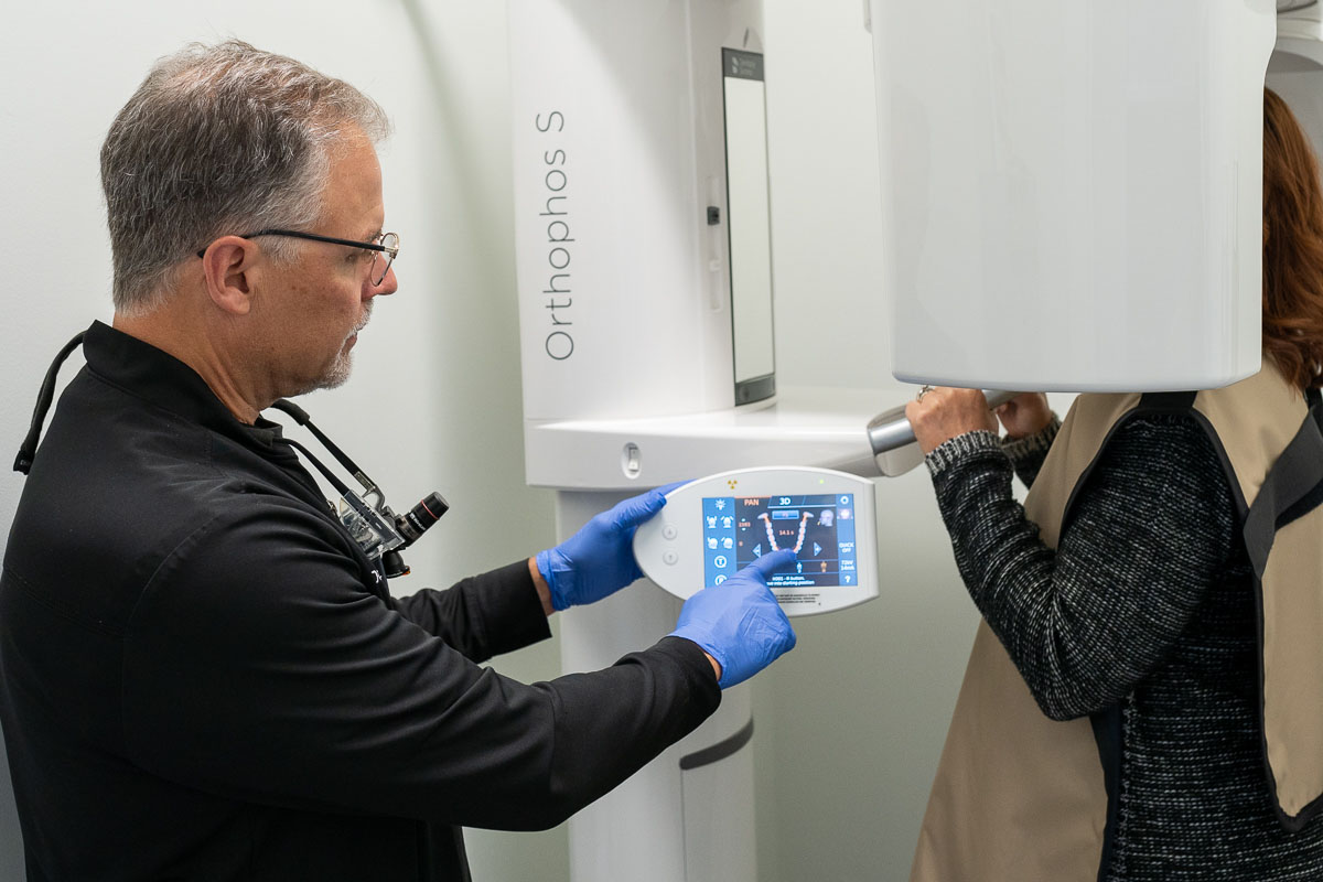
- Sirona Orthophos SL 3D panorex and cone beam system features panoramic and selectable 3D imaging based on the individual diagnostic needs. It offers a range of panoramic exams, 3D field of views for endodontics or single tooth implants, dual jaw or full arch implants and the ability for Dr. Michelakis to view “down through” any tooth or section of the jaw.
Radiographs allow us to see everything we cannot see with our eyes to detect cavities in between your teeth, determine bone level, and analyze the health of your bone. We can also examine the roots and nerves of teeth, diagnose lesions such as cysts, infections or tumors, as well as assess damage when trauma occurs.

DIAGNOdent LASER DECAY SENSOR
The DIAGNOdent uses laser fluorescence to aid in the detection of caries within the tooth structure. As the incident laser light is dispersed into the site, carious tooth structure will exhibit fluorescence, proportionate to the degree of caries, resulting in audible elevated scale readings on the display. Clean healthy tooth structure exhibits little or no sound or fluorescence and will result in very low scale readings on the display.

HIGH RESOLUTION INTRA-ORAL CAMERA
The Sopro 717 Intra-oral is a fixed-focus camera that offers instant, adjustment-free focusing with a great depth of field, providing more that 100x magnification to enhance Dr. Michelakis’ ability to diagnose and treat and areas of decay or periodontal conditions. The camera itself is no bigger than an electric toothbrush allowing for detailed photos of all the teeth and supporting structures.

CEREC MILLING UNIT
CEREC® is a registered trademark of Sirona Dental Systems
CEREC
CEREC is a method used by thousands of dentists worldwide since 1987 not only to replace fillings and/or crowns, but also to restore any tooth that is decayed, weakened or broken, to its natural strength and beauty.
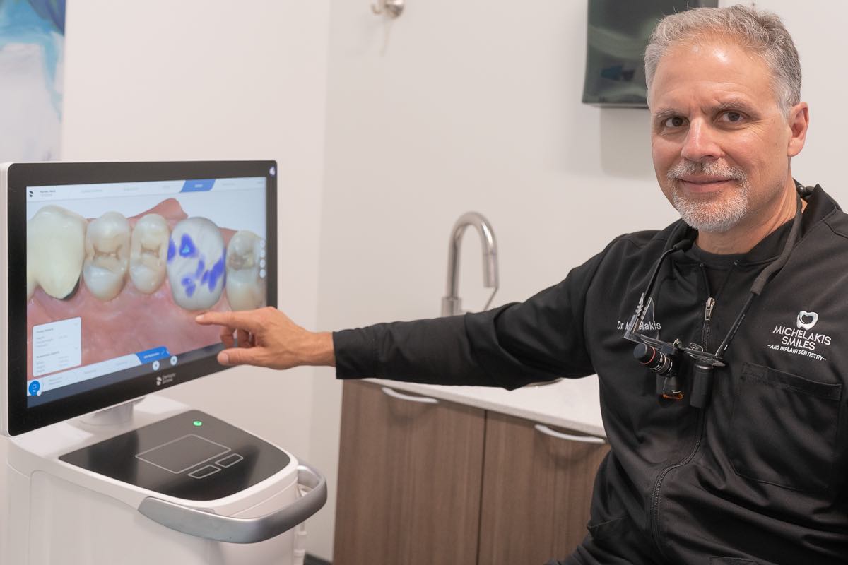
CEREC Primescan Scanner Unit
The CEREC Primescan Scanner Unit is a computer, scanner and camera. The camera to takes a digital picture of your prepared tooth. This picture is used instead of a traditional impression. This means no impression tray and material for you to gag on. The computer and CEREC 3D software convert the digital picture to a three-dimensional virtual model of your tooth.
Dr. Michelakis then designs your restoration right on screen using the software while you wait (and watch!). This software assists Dr. Michelakis with designing any single tooth restoration: crowns, inlays (fillings), onlays (partial crowns) and bridges. Once Dr. Michelakis has designed your restoration (usually about 5 minutes), he clicks a button, and the design data is sent to the CEREC Milling Unit here in the office.
CEREC milling unit
The CEREC milling unit creates your restoration. The CEREC 3D software takes the digital images and converts it into a 3-dimensional virtual model on the computer screen. A ceramic block that matches your tooth shade is placed in the milling machine. The data from the Acquisition Unit is used to direct two diamond-coated burs to carve the block into the exact shape of the restoration and/or tooth crown.